Article Text

Abstract
Background: The gene encoding fatty acid CoA ligase 4 (FACL4) is mutated in families with non-specific X linked mental retardation (MRX) and is responsible for cognitive impairment in the contiguous gene syndrome ATS-MR (Alport syndrome and mental retardation), mapped to Xq22.3. This finding makes this gene a good candidate for other mental retardation disorders mapping in this region.
Methods: We have screened the FACL4 gene in eight families, two MRX and six syndromic X linked mental retardation (MRXS), mapping in a large interval encompassing Xq22.3.
Results: We have found a missense mutation in MRX68. The mutation (c.1001C>T in the brain isoform) cosegregates with the disease and changes a highly conserved proline into a leucine (p.P375L) in the first luciferase domain, which markedly reduces the enzymatic activity. Furthermore, all heterozygous females showed completely skewed X inactivation in blood leucocytes, as happens in all reported females with other FACL4 point mutations or deletions.
Conclusions: Since the FACL4 gene is highly expressed in brain, where it encodes a brain specific isoform, and is located in hippocampal and cerebellar neurones, a role for this gene in cognitive processes can be expected. Here we report the third MRX family with a FACL4 mutation and describe the development of a rapid enzymatic assay on peripheral blood that we propose as a sensitive, robust, and efficient diagnostic tool in mentally retarded males.
- non-specific X linked mental retardation
- FACL4
- MRX68
Statistics from Altmetric.com
Mental retardation is a condition characterised by significantly below average intellectual functioning (IQ<70) in conjunction with significant limitations in adaptive functioning, occurring before the age of 18 years.1 An excess of males in mentally retarded patients has long been noticed and explained by the presence of mutations in one gene on the X chromosome.2 It is calculated that X linked mental retardation (XLMR) may account for about 20–25% of mentally retarded males.3–5 Usually, XLMR is divided into “syndromic” (MRXS) and “non-specific” (MRX) forms, depending on the presence or absence, besides mental retardation, of a specific pattern of physical, neurological, or metabolic abnormalities. Up to now, 11 genes have been found to be involved in MRX. This number of genes may rise to more than 100 since the mutation frequency of the identified genes is very low (0.01–1%) and no major MRX gene seems to exist.6 For several of the reported genes, knowledge of the pathogenesis is very limited and apparently they do not seem to participate in a common pathway: FMR2 on Xq28 encodes a nuclear protein that may be a transcriptional regulator7,8; GDI1, on Xq28, is implicated in synaptic vesicle cycling and neurotransmitter release9; TM4SF2 (alias MXS1), on Xp11.4, interacts with beta-1 integrins and may have a role in the control of neurite outgrowth10; IL1RAPL1, on Xp22.1-Xp21.3, is related to interleukin-1 receptor accessory proteins.11 Three genes, the oligophrenin-1 gene (OPHN1) on Xq12, PAK3 on Xq21.3-q24, and ARHGEF on Xq26, encode proteins involved in the Rho GTPases pathways.12–15 These signal transduction pathways are thought to mediate organisation of the cytoskeleton, cell shape, and motility, and may be responsible for axonal outgrowth and the shape and size of dendrites. Three genes, RPS6KA3 (RSK2) on Xp22, MECP2 on Xq28, and ARX on Xp21.3–22.1, are associated with both MRX and MRXS (Coffin-Lowry, Rett, X linked West, and Partington syndromes). These three genes may be involved in a common pathway of gene expression regulation and chromatin remodelling, indicating that the disruption of another cellular process, beside the Rho-GTPases cycle, may lead to MR.16,17,18,19
The human long chain fatty acyl-CoA ligase (or synthetase) 4 (FACL4 or (L)ACS4) gene on Xq22.3 may be involved in a new MR pathogenic pathway owing to alteration of lipid metabolism.20 We first reported that this gene is included in a large genomic deletion leading to a contiguous gene syndrome characterised by Alport syndrome, mental retardation, midface hypoplasia, and elliptocytosis (AMME, OMIM 300194).21,22 Subsequently, we showed that the same gene is also deleted in a second family with a smaller genomic deletion leading to Alport syndrome and mental retardation and we proposed to rename the contiguous gene syndrome ATS-MR.23 Finally, we confirmed that the FACL4 gene is indeed involved in MR through the identification of a missense mutation (p.R529S) in the FACS signature motif of the protein in MRX63, and of one splice site mutation in a family without linkage data.20 Both these mutations abrogate the arachidonyl-CoA synthetase activity, and this abrogation might disturb an important developmental lipid associated cognitive process. The present study describes a third MRX family with a novel mutation in the FACL4 gene and proposes a rapid and sensitive assay to screen for FACL4 enzymatic activity in blood samples.
SUBJECTS AND METHODS
MRX68
The pedigree of family MRX68 (proband L46) includes five affected males, one of whom (II.5) died at 8 months of age. At the time of this study, four affected males (II.6, II.7, III.2, and III.3) were still alive, and all were available for clinical examination and blood sampling.
To determine the level of intelligence two domains were explored and included measurements of the mental level (including behaviour) and adaptive functioning.24 To assess the mental level, cognitive strengths and weaknesses, the Wechsler Intelligence Scale for Children Revised (WISC-R)25 was used in II.7 and III.3, and the Wechsler Adult Intelligence Scale (WAIS)26 in II.1, II.3, II.8, III.1, and III.2. Behavioural observations were done with the “Child Behavior Check List (CBCL)” in III.3 in combination with the “Teacher’s Report Form (TRF)”. The “Youth Self Report (YSR)”27 was obtained from III.2 and III.3. To measure the level of adaptive functioning, the “Assessment of Adaptive Areas (AAA)” was used.28 The Dutch versions of all these tests and checklists were used.
All affected family members were subjected to physical examination, including biometric measurements, neurological, and clinical genetic investigations (table 1).
Biometric measurements and cognitive levels in the MRX68 family
Other investigated families
In addition to family MRX68, we examined seven families showing linkage to large intervals of the X chromosome covering Xq22.3 collected by Leuven (probands L22, L49, L56) and by the Greenwood Genetic Center, South Carolina (families K8045, K8435, K8835, K8610).
Proband L22 belongs to family MRX35. The males have a non-specific condition with severe hyperkinetic behaviour and moderate mental retardation. Two point and multipoint linkage analysis with 18 highly polymorphic markers covering the entire X chromosome assigned the mutant locus to the Xq22-q26 region.29
Probands L49 and L56 belong to two families with mental retardation and spastic paraplegia, mapping in Xp11.4-Xq24 (and Xq27-Xq28) and Xp11.3-Xq24, respectively (XLMR meeting, Strasbourg 1999 (L56) and Frascati 2001 (L49)30).
Family K8435 has moderate mental retardation. Affected males have distinct facial features, hirsutism, seizures, unsteady gait, and slowly progressive weakness. Most affected males have hypogammaglobulinaemia with specific absence of plasma and/or secretory IgA.31 Two point linkage analysis, using 27 microsatellite markers on the X chromosome, places the mutant locus between Xq21.33 and Xq23.
All affected males of family K8045 had a normal facial appearance, moderate to severe MR and spastic paraplegia and they had never walked.32 A seven point linkage analysis yielded a maximum lod score for three loci spanning Xq22-q25.
In family K8610, males have agenesis/hypoplasia of the corpus callosum and features of FG syndrome.33 Linkage analysis assigned the locus to Xq12-Xq22.3.
Family K8835 is unpublished. All affected males have significant developmental delay with variable phenotypic presentation. Macrocephaly, unusual gait, and macro-orchidism are variously combined in the affected males of the family. Moreover, two of them have a prominent forehead and one has a round face. They are FRAXA negative. Linkage analysis placed the mutant locus on Xp11.3-Xq23.
Linkage analysis
Highly polymorphic microsatellite markers spread over the entire X chromosome were analysed on genomic DNA extracted from peripheral white blood cells of all family members. PCR amplification of these polymorphic markers was performed using fluorescein labelled locus specific primer pairs (Genome Database). Marker genotypes were determined by separation of PCR products on an ALF sequencer (Amersham Pharmacia Biotech). Sizing of the PCR products was done with the Allelelinks software (Amersham Pharmacia Biotech). Multipoint lod score analyses were performed using MLINK version 5.134 and FASTLINK version 2.2.35 Map locations, genetic distances, and allele frequencies were obtained from the Genome Database (http://gdbwww.gdb.org/), the Location Database (http://cedar.genetics.soton.ac.uk/pub/chromX/gmap), Ensembl (http://www.ensembl.org), and Généthon data.36 The mutation rate was set at 3 × 10⁻⁶ and gene frequency at 0.0001. For males, the penetrance was set at 0.0 and 1.0 for non-carriers and carriers of the disease gene, respectively. For females, the penetrance was set at 0.0 for non-carriers, 0.25 for heterozygous carriers, and 1.0 for homozygous carriers of the disease gene.
X inactivation studies and mutation analysis
X inactivation studies in all females, including female carriers of family MRX68 (I.2, II.3, and II.8), were performed using the previously described protocol37 on DNA extracted from peripheral blood. Mutation analysis of the FMR1 gene (the most common cause of XLMR) was performed using standard procedures.38,39 The FACL4 coding region, encompassing exons 3–17 and covering both alternative start codons located in exons 3 and 4, respectively, was PCR amplified with specific primers.20 Direct sequencing of PCR products was carried out in both directions with BigDye Terminator Cycle Sequencing Kit (PE Applied Biosystems, Foster City, CA) on an ABI 310 Automated Sequencer. Sequences were analysed with the Genescan package software.
Segregation analysis in family MRX68 for the novel mutation identified in exon 10 was performed by MspI digestion of the PCR product of this exon (primers: F: 5′-AATTCCAAGTGTAACTTCTG-3′ and R: 5′ TAAAAGGTCCAA GTACGATC-3′). The digest was analysed on a 1.5% agarose gel. Loss of the MspI site was confirmed by direct sequencing.
Enzymatic assay
To test FACL4 enzymatic activity in whole cell lysates obtained from EBV transformed cell lines, we have modified our original method20 as follows: 107 cells, instead of the original 108, were harvested and resuspended in 200 μl of lysis buffer. Before proceeding with the test, 20 μl of the lysate were removed from each sample and used for protein quantification using the BIO-RAD protein assay (BIO-RAD). The remaining cell lysate was incubated for 20 minutes in 150 μl of a standard reaction mixture (100 mmol/l TRIS/HCl, pH 8.0, 6.67 mmol/l ATP, 0.66 mmol/l CoA, 5 mmol/l dithiothreitol, 20 mmol/l MgCl2), and 40 μl of a solution containing arachidonic acid (50 mmol/l NaHCO3, 7.5 mmol/l Triton X-100, 0.25 mmol/l arachidonic acid, corresponding to 10 nmol, and 2 × 10⁵ dpm of 14C labelled arachidonic acid). The reaction was stopped with 2.25 ml of 2-propanol:heptane:2 mol/l sulphuric acid (40:10:1), followed by 1.5 ml of heptane and 1 ml of water and vigorous shaking. After centrifugation (five minutes at 2000 rpm), the upper layer was removed and the lower aqueous phase was extracted three times with 2 ml of heptane. The radioactivity in the upper (heptane) layers, containing arachidonic acid, and in the lower (aqueous) phase, containing arachidonyl-CoA, was determined by scintillation counting (Beckman). To determine enzyme activity, the total radioactivity (upper plus lower phase) and the percentage of this radioactivity in the lower phase were calculated. This percentage corresponds to the percentage of arachidonic acid used for the reaction (10 nmol) which has been converted to arachidonyl-CoA. The values were corrected for protein quantity. FACL4 enzymatic activity was expressed as nmol of arachidonyl-CoA obtained per mg of total protein.
To perform the test in blood samples, 10 ml of blood was diluted with one volume of phosphate buffered saline (PBS) or physiological solution, mixed, and carefully layered on one volume of a solution containing Ficoll 99 g/l, sodium chloride 12 mmol/l, and sodium diatrizoate 0.16 mol/l. After centrifugation (2000 rpm for 40 minutes at room temperature), the upper layer of plasma and platelets was removed and the intermediate layer containing leucocytes was recovered into a fresh tube and washed twice with PBS or physiological solution. In order to eliminate the residual erythrocytes present after the treatment with Ficoll, the pellet of leucocytes was resuspended in 1 ml of water, incubated in ice for one minute, and then diluted to 10 ml with PBS or physiological solution and centrifuged at 2000 rpm for 10 minutes. Leucocytes were also isolated from blood samples conserved at room temperature for 24, 72, and 120 hours.
For cryopreservation, leucocytes were isolated from 10 ml of fresh blood using the Ficoll method as described above and then stored at −80°C until the test was performed. Both cryopreserved and room temperature conserved leucocytes were subjected to the enzymatic test using the protocol described above.
RESULTS
Molecular analysis
We have screened by direct sequencing the whole coding region of FACL4 in eight X linked mental retardation families, two non-specific (MRX35 and MRX68), and six syndromic, mapping in a large region encompassing Xq22.3.29,31–33,40 A FACL4 pathogenic mutation was found in family MRX68. Multipoint linkage analysis, previously performed on this family using 25 highly polymorphic markers, indicated a candidate gene region of 12.5 cM between markers DXS8020 (Xq22.1) and DXS1220 (Xq23). A maximum lod score of 2.1 was obtained at markers DXS1153 and COL4A5 (fig 1).

Multipoint lod score analysis of family MRX68.
Mutation analysis of the FACL4 gene in the family showed a missense mutation, c.1001C>T (brain isoform), which changes a highly conserved proline (CCG) at amino acid position 375 into a leucine (CTG) (figs 2 and 3). This resulted in the abrogation of a restriction site for MspI. Restriction analysis showed the presence of the mutation in all affected males and carrier females (fig 4). Sequence analysis confirmed the C>T transition in these people. The mutation was not found in 300 normal chromosomes.

Chromatogram of mutated sequence. Sequence analysis showed a C>T transition at cDNA position 1001, changing a proline (CCG) into a leucine (CTG). Both the nucleotide and amino acid change are shown above the chromatogram. Intron bases are indicated in lower case letters. An arrow indicates the mutated nucleotide. Wt = wild type sequence. M = mutated sequence.

Schematic representation of FACL4 brain specific isoform. The p.P375L substitution in MRX68 at the end of the first luciferase domain is indicated. The mutations previously found in other families are also shown. LR1 = luciferase domain 1, LR2 = luciferase domain 2, striped box = adenosine monophosphate binding motif, grey box = FACS consensus sequence, large circle = brain specific amino terminal peptide (41 amino acids). Zig zag line = truncation of the protein by the splice site mutation, c.1003-2 A>G. Small circles = missense mutations. Numbers at the top refer to the amino acid positions.
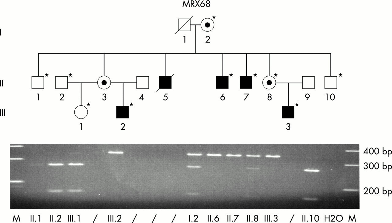
Segregation analysis. The DNA (*) of members of family MRX68 was analysed on an agarose gel after MspI (CCGG) digestion of the PCR product of FACL4 exon 10. The site was lost in all affected males. A heterozygous loss of the site was observed in the obligate carrier females. M = molecular weight marker with sizes indicated on the right. A slash (/) indicates empty lines. H2O = negative PCR control without DNA.
The pedigree and clinical findings in the family are most compatible with the diagnosis of MRX (table 1). The affected males did not show facial dysmorphism or neurological abnormalities. An MRI scan of one affected subject was normal (data not shown). The neurocognitive levels ranged from mild (II.7) to moderate (III.2, III.3) MR in the affected males and the intelligence levels were borderline in female carriers (II.3, II.8) (table 1). The personality profile was analysed in three affected males (II.7, III.2, III.3) and all obtained poor results on the dimensions “motor activity” and “creativity”. Internalising behavioural problems were observed in one subject (III.3), together with severe social problems, thought problems, and attention deficit. Autistic-like features were present in this child.
X inactivation studies in the obligate carrier females of the family (I.2, II.3, and II.8) showed 100% skewed inactivation profiles in all of them (data not shown).
Enzymatic assay
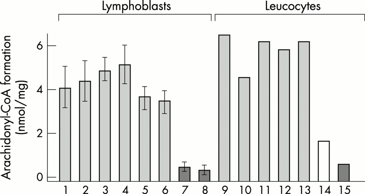
Analysis of the arachidonyl-CoA synthetase activity was performed on whole cell lysates obtained from both an EBV transformed cell line of an affected male (II.7) and leucocytes isolated from fresh blood of the same male. In both cases the analysis showed a marked reduction in activity in the patient with respect to normal controls (fig 5, columns 7 and 15). The reduction in activity was of the same extent as that observed in a male with FACL4 deletion,20 indicating that the p.P376L mutation strongly inhibits FACL4 activity (fig 5, column 8).
Arachidonyl Co-A synthetase activity. The assay was carried out on whole cell lysates of 107 lymphoblastoid cell lines (columns 1–8) and of leucocytes isolated from 10 ml of blood samples (columns 9–15). For lymphoblastoid cell lines, results show the means of at least three independent experiments. Statistical evaluation between groups was done with Student’s t test (p=0.01). Column 1 = normal control; 2 = L22; 3 = K8045; 4 = K8435; 5 = K8835; 6 = K8610; 7 = L46 (MRX68); 8 = male patient with genomic deletion (ATS-MR syndrome); 9 = L49; 10 = L56; 11 = control from 10 ml of blood after cryopreservation. Lanes 12–14 = controls after 24, 72, and 120 hours of blood preservation at room temperature; 15 = blood of L46 (MRX68) stored at room temperature for 24 hours. Columns 7, 8, and 15 (dark grey box) and column 14 (white box) were significantly different from controls.
In order to exclude FACL4 intronic/promoter mutations, missed by coding sequence analysis, FACL4 enzymatic activity was tested also in lymphoblastoid cell lines or leucocytes of each proband of the other seven families. No statistical differences were found in the arachidonyl-CoA synthetase activity between these probands (fig 5, columns 2–6 and 9–10) and controls.
Assessment of enzymatic assay on blood
When a disease gene encodes an enzyme expressed in leucocytes, a functional assay could be proposed not only to confirm a mutation but also to replace molecular analysis as a screening method. However, in our original description the method required the EBV transformation of B lymphocytes. This step is time consuming and expensive. With the aim of bypassing the cell transformation step and to apply the test directly to fresh blood, we have gradually reduced the number of cells used for the test from 108 to 107. This number corresponds to the mean amount of leucocytes present in 10 ml of blood. Our analysis showed that 107 cells are enough to detect arachidonyl-CoA synthetase activity and to distinguish a FACL4 mutation (fig 5, columns 1–8). In addition, enzymatic activities observed with 107 lymphoblastoid cells and with leucocytes isolated from 10 ml of blood are comparable both in patients and in controls (fig 5, columns 1–8 v 9–15). Moreover, we tested whether blood may be cryopreserved or stored at room temperature for several days before performing the analysis. Fig 5 shows that there is no difference in activity after cryopreservation (column 11) or after 24 or 72 hours of conservation at room temperature (columns 12–13). A significant reduction in activity (p=0.01) was noted after 120 hours at room temperature (fig 5, column 14).
DISCUSSION
We performed mutation analysis of the FACL4 gene in family MRX68 and in seven other families that map in large regions encompassing Xq22.3. In MRX68 a novel mutation was identified that causes the substitution of a highly conserved proline with a leucine (p.P375L in brain isoform). The FACL4 protein consists of two luciferase domains, the precise functions of which remain to be elucidated (fig 3). The conserved FACS signature motif, located in the second luciferase domain, is thought to be important for the binding of fatty acids and hence should play a crucial role in lipid metabolism. The recently reported missense mutation in MRX63 changes a conserved arginine within the FACS motif into a leucine, thereby interfering with the enzymatic activity.20 In the same study, a mutation in intron 10, identified in another small family, uncovered a cryptic splice site that introduced a premature stop codon. This mutation is predicted to lead to a protein lacking the second luciferase domain including the FACS signature motif. The new missense mutation presented here (p.P375L) is located at the end of the first luciferase domain that contains a putative AMP-binding signature motif. The mutated proline is highly conserved between all members of the FACL family including those of other species, and it can be hypothesised that sequence similarities presumably reflect functional requirements that are common to these enzymes. Therefore, this novel missense mutation is likely to be pathogenic since it cosegregates with the disease, strongly inhibits FACL4 activity, involves a highly conserved residue, and was not found in 300 control chromosomes. Fatty acid binding assays and structural models of the wild type and mutant proteins will help to determine the precise function of the amino acids mutated in this and in the other families.
In the last few years, pathogenic mutations in the MECP2, RPS6KA3, and ARX genes have been identified in patients with both syndromic (MRXS) and non-specific (MRX) X linked mental retardation, indicating that the same gene can be responsible for both conditions.16–19 Given this, in addition to the two MRX families, we also analysed for FACL4 mutations six MRXS families in which linkage analysis assigned the mutant locus to regions encompassing Xq22.3, suggesting a possible involvement of FACL4. No mutations were identified in these families, excluding the involvement of FACL4 in their pathogenesis. However, given the small number of families tested, no conclusions regarding the role of FACL4 in MRXS can be drawn. A larger panel of patients needs to be analysed in order firmly to establish whether FACL4 is also involved in the onset of MRXS.
Long chain fatty acyl-CoA synthetases convert free fatty acids into fatty acyl-CoA esters, which are the key intermediates in the synthesis of complex cellular lipids (triglycerides, phospholipids, and cholesterol esters).41 The highly preferred substrates for FACL4 include arachidonic acid (AA) and eicosapentaenoic acid (EPA).42 Compared to other tissues, brain tissue contains high amounts of polyunsaturated fatty acids (PUFAs), such as AA, EPA, and docosahexaenoic acids (DHA) in its membrane phospholipids. The central nervous system uses these PUFAs when cellular differentiation, active synaptogenesis, and photoreceptor membrane biogenesis takes place.43 AA and DHA are the most abundant long chain PUFAs in brain, and influence membrane fluidity, recovery from injury, gene transcription, and signal transduction via protein kinase C, in a Ca2+ and diacylglycerol dependent way.44–47 Long chain acyl CoA esters have been reported to affect a large number of cellular processes including ion distribution, enzyme regulation, vesicle transport and membrane fusion, and gene expression.48 This is particularly relevant when considering the possible effect of arachidonyl-CoA deficiency on neural development. Moreover, several randomised controlled studies show that long chain PUFAs do affect infant cognitive development, especially visual attention and problem solving.43 Finally, it has been shown that EPA might prevent interleukin-1β and radiation induced deterioration in rat neurones.49,50 Therefore, considerable reduction of the enzymatic activity of FACL4 in the brain might lead to deranged fatty acid metabolism in neurones causing defects of neurone outgrowth, synaptogenesis, or other developmental restrictions important for normal brain development.
The functional assay of FACL4 activity is a direct method to test whether the FACL4 gene may be involved in a mentally retarded patient. We have modified the original enzymatic assay, which required EBV transformation of lymphocytes, into a simple test on blood and we propose this test for rapid screening of mentally retarded males. This enzymatic approach has a number of advantages over the standard molecular analysis and it is less laborious, much faster, and reagent intensive. On the basis of our experience, the operational cost of FACL4 analysis by this enzymatic strategy is 75% lower than mutation analysis performed by either mutation screening methods or full sequencing. In addition to the increased cost efficiency, the diagnostic enzymatic strategy has proven to be a sensitive and efficient method for FACL4 investigation. Moreover, this approach allows the identification of functionally harmful intronic and promoter mutations that are usually missed by standard mutation analysis of coding sequences. Finally, by direct measurement of enzymatic activity, this method overcomes interpretation uncertainty commonly associated with missense changes. We showed that the test can be performed in blood conserved at room temperature up to 72 hours and thus can be applied to shipped samples from anywhere.
In conclusion, we describe a novel missense mutation in the FACL4 gene in a family with non-specific mental retardation. The p.P375L mutation results in a big reduction of enzymatic activity, which might disturb cognition related brain lipid metabolism. Our data confirm the involvement of FACL4 in MRX. Moreover, we developed a rapid enzymatic assay on peripheral blood which we propose as a sensitive, robust, and efficient diagnostic tool in mentally retarded males. This is the first biochemical assay that can be used to identify mutations in mentally retarded patients.
Acknowledgments
We thank the family members for their participation and assistance, Philippe Volcke and Lut van den Berghe, Connie Schrander-Stumpel and Christine de Die for their expert clinical support, and Marianne Wouters for psychological testing. This work was supported by a Telethon grant (E.1145) to AR and in part by the Fund for Scientific Research of Flanders (Belgium) grant FWO-G0182.97 and G0229.01 (A5334).