Article Text

Statistics from Altmetric.com
Microcephaly is the clinical finding of a head circumference measurement greater than three standard deviations (SD) below the population mean for age and sex. It is usually accompanied by mental retardation and there are many diagnoses with both environmental and genetic aetiologies.1 Autosomal recessive primary microcephaly (MCPH) (MIM 251200) is a disorder in which affected subjects are born with a small head circumference, explained by a cerebral cortex of reduced size, and are mentally retarded. The brain is structurally normal and, apart from the intellectual impairment, there are no other significant neurological problems, dysmorphic features, or malformations.2,3 In a study carried out in The Netherlands,4 the incidence of MCPH was approximately 1/250 000 but it is probably greater in populations with a high rate of consanguineous marriages. MCPH has been shown to be genetically heterogeneous with the identification of five loci: MCPH1 on 8p23,5 MCPH2 on 19q13,6 MCPH3 on 9q34,7 MCPH4 on 15q15-q21,8 and MCPH5 on 1q31.9,10 MCPH1, 2, and 3 were mapped in northern Pakistani families, MCPH4 in a Moroccan family, and MCPH5 in northern Pakistani and Turkish families. Here we report the identification by autozygosity mapping11 of a novel locus for primary microcephaly, MCPH6, in a north eastern Brazilian family.
Key points
-
Autosomal recessive primary microcephaly (MCPH) is a genetic disorder in which an affected subject is born with a head circumference >3 SD below the expected mean and is mentally retarded.
-
We report a novel locus (MCPH6) mapped to chromosome 13q12.2 in a Brazilian family.
-
The minimal critical region spans 6 Mb between markers AL139378GT17 and D13S1244 with a maximum two point lod score of 6.25.
MATERIALS AND METHODS
Subjects
The consanguineous family had eight affected subjects (five males and three females, DNA available from seven subjects), with ages varying between 4 and 27 years (fig 1), in four sibships (fig 2). The head circumference of all affected subjects was noted to be small at birth and between 7-10 SD below the expected mean when examined by us. All had mental retardation of moderate severity: the three adults and the adolescent affected were unable to read or write but could speak simple phrases and had basic self-care skills. With the exception of intellectual impairment, there were no other neurological problems (including fits) and motor development had been normal. All eight were in good health and had growth parameters within normal limits. They were not dysmorphic and no syndrome diagnosis could be made. No past medical history or environmental causes could be found to explain the finding of microcephaly. The parents had normal head circumference and intelligence. Ophthalmological examination, standard lymphocyte karyotype (400 bands), and electroencephalogram performed in four affected subjects were normal, and brain scans in two showed no cerebral malformations or neuronal ectopia.

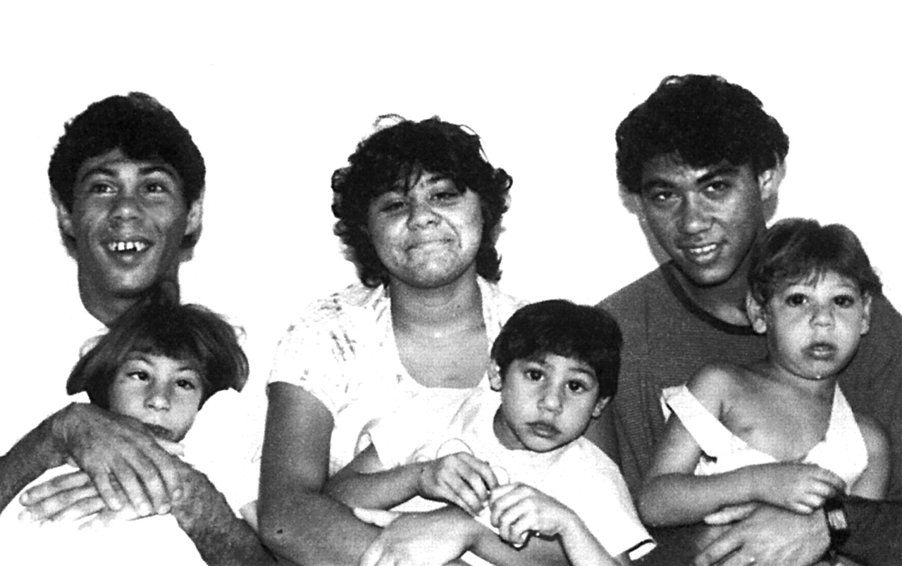
Six of the eight affected subjects with ages between 4 and 27 years with a diagnosis of autosomal recessive primary microcephaly.

Genotypes for eight markers used in the study at 13q12.2 arranged centromere to qter. Unaffected sibs have been omitted for clarity. Marker order was taken from the Marshfield linkage map. The boxed region shows the shared region of homozygosity in affected subjects. The FGF9 gene, indicated by an arrow, is flanked by markers AL139378GT21 and AL139378GT17 and hence is excluded as a candidate gene from the common homozygous region in affected subjects.
Molecular genetics
Linkage to the five known MCPH loci was ruled out (data not shown). An autosomal chromosome screen for regions of shared homozygosity was performed on seven of the eight affected subjects and their parents with the CHLC/Weber Human Screening Set version 8 (Research Genetics), which contains 365 autosomal microsatellite repeat markers spaced at approximately 10 cM intervals. PCR amplification of all markers was performed according to the manufacturer’s specifications using a Roboseq 4200 (MWG BioTech Ltd). Amplified markers were pooled and electrophoresed on an ABI Prism 377 gene sequencer (Applied Biosystems) on 4.2% polyacrylamide gels, at 3000 V and 52°C, for 2.5 hours. Fragment length analysis was undertaken using the ABI Prism Genescan and Genotyper 1.1.1 analysis packages.
RESULTS
A single region of homozygosity common to all seven microcephalic subjects was identified on chromosome 13q defined by markers D13S787 and D13S1304. Further refinement of the region was conducted using the following markers selected from the ABI Linkage Mapping Panel Version I (Applied Biosystems), the Todd Panel,12 and the Marshfield Linkage Maps: cen - D13S175 - D13S1275 - D13S787 - D13S221 - D13S1304 - D13S1254 - D13S1244 - D13S217 - D13S120 - D13S171 - D13S1493 - tel. This defined a shared homozygous region on chromosome 13 at band q12.2 with meiotic crossovers between markers D13S175-D13S1275 and D13S1254-D13S1244, with the centromeric and telomeric boundaries of a 9 cM region being defined by D13S175 and D13S1244. Information regarding marker order and relative distances was obtained from the Marshfield Linkage Maps. The marker order obtained from the Marshfield Linkage Maps was in agreement with that derived from analysis of the current draft human genome data.
A fully penetrant autosomal recessive mode of inheritance and a disease gene frequency of 0.003 were assumed. Owing to the complexity of the family structure, equal allele frequencies were assumed for each marker when calculating the lod scores and the maximum number of alleles was set at 4. Pedigree allele inconsistencies were identified using PedCheck.13 Two point analysis was performed using the LINKAGE analysis programs14 at θ=0 for markers in the critical region and results are shown in table 1 with the highest lod score at 6.25 for marker D13S1275.
Two point lod scores at θ=0 for each marker defining the MCPH6 region at 13q12.2
Novel microsatellite markers to refine the region further were designed using the Human Genome Browser and the Primer3 program, and designated [human BAC accession number][microsatellite repeat unit][number of unit repeats in the reference BAC], for example, AL356285TG25 (fig 2). These allowed us to redefine the centromeric boundary marker as AL139378GT17.
DISCUSSION
Haplotype and lod score analysis both suggest that the chromosome region 13q12.2, designated the MCPH6 locus, contains a gene which when mutated causes autosomal recessive primary microcephaly.
Within the larger MCPH6 region of 9 cM there is the potential candidate gene, fibroblast growth factor 9 (FGF9). In the nervous system of mice, FGF9 is produced mainly by neurones and may have a role in glial cell growth and differentiation during development.15,16 The redefinition of the region to 6 cM using novel microsatellite markers flanking FGF9 resulted in the exclusion of this gene (fig 2). We now therefore consider that the gene causing this form of autosomal recessive primary microcephaly must lie within this smaller region of approximately 6 Mb. To date, only the MCPH1 gene, microcephalin, and the MCPH5 gene, ASPM, have been identified.17,18 Future identification of the MCPH6 gene may be aided by an insight into how these proteins function and interact within the human brain, such as mitotic spindle activity in the case of ASPM. The discovery of MCPH genes will lead to a greater understanding of normal and abnormal human fetal cerebral cortex growth, giving potential insights into the question of how the mammalian cerebral cortex evolved and has become so predominant in humans, and the wherewithal to offer diagnostic, prenatal, and carrier testing for affected families.
Acknowledgments
We express our gratitude to the members of the family studied. This work has been funded by CAPES (Coordenação de Aperfeiçoamento de Pessoal de Nível Superior), FACEPE (Fundação de Amparo à Ciência e Tecnologia do Estado de Pernambuco), the Wellcome Trust, and the West Riding Medical Research Trust. We thank the Research Center Aggeu Magalhães (CPqAM) for permitting us to use their equipment for DNA extraction and the staff of HGMP for computational assistance.
Electronic Database Information. URLs for data in this article are as follows: Center for Medical Genetics, Marshfield Medical Research Foundation available at http://research.marshfieldclinic.org/genetics/ (for genetic linkage maps). Online Mendelian Inheritance in Man (OMIM): http://www.ncbi.nlm.nih.gov/omim. Human Gene Nomenclature Database, HUGO Gene Nomenclature Committee: http://www.gene.ucl.ac.uk/nomenclature/ Draft human genome browser: http://genome.cse.ucsc.edu/. For primer creation (for novel microsatellite markers): http://www-genome.wi.mit.edu/cgi-bin/primer/primer3_www.cgi.