
JNeurosci publishes a broad spectrum of articles reporting important work across areas of neuroscience. Because keeping up with exciting work outside your individual subdiscipline can be difficult, JNeurosci created the Spotlight feature to highlight articles that our reviewers gave the highest marks for both methodological merit and significance. We hope the papers in this year’s Spotlight will be of interest to neuroscientists across areas and at many different levels of experience. Below is the collection of this year’s Spotlight papers and summaries of their key findings. Papers selected for the Spotlight feature in previous years can be found below as well.
2023

Behavioral/Cognitive
Structural Fingerprinting of the Frontal Aslant Tract: Predicting Cognitive Control Capacity and Obsessive-Compulsive Symptoms
The frontal aslant tract allows communication between brain regions that regulate the processing of new information and the control of reflexive behaviors. The connectivity of these regions is thought to be uniquely individual, but the individuality of the frontal aslant tract and its role in cognitive control were unknown prior to this rigorous human imaging study by Wang and colleagues. They used multiple datasets and complementary analyses to discover that the frontal aslant tract is a structural fingerprint of the brain capable of predicting cognitive control and obsessive-compulsive disorder severity.

Behavioral/Cognitive
Spontaneous α Brain Dynamics Track the Episodic “When”
A major component of our understanding of reality is the feeling that time passes, which requires tracking the passage of time. A century-old working hypothesis suggests our “internal clocks” are driven by brain activity called “alpha” rhythms. Azizi et al. explored this link more directly by using magnetoencephalography to record from human brains under two time-tracking behavioral conditions. They discovered that the bursting time of alpha rhythms reliably indicated how much time an individual would report to having elapsed, but that alpha wave time tracking vanished when individuals predicted the amount of time and were attending to it during recording. This novel link between oscillatory bursts and spontaneous cognition may shape the direction of study for this field of research.

Cellular/Molecular
Selective Serotonin Reuptake Inhibitors within Cells: Temporal Resolution in Cytoplasm, Endoplasmic Reticulum, and Membrane
Selective serotonin reuptake inhibitors (SSRIs), such as fluoxetine and escitalopram, are given to millions of people for mood stabilization. It is assumed that these therapeutic drugs increase extracellular serotonin levels by inhibiting the transporter that clears serotonin from synapses. However, the biophysical mechanism of SSRIs may be more complex than that. In this study, Nichols et al. developed novel tools to investigate the accumulation and kinetics of fluoxetine and escitalopram in different cellular compartments, using both primary neurons and neuronal cell lines for their observations. Their findings may inform future exploration on where and how SSRIs engage therapeutic targets.

Cellular/Molecular
GRM2 Regulates Functional Integration of Adult-Born DGCs by Paradoxically Modulating MEK/ERK1/2 Pathway
Dentate granule cells (DGCs) that develop in adulthood form hippocampal neural circuits that serve major roles in regulating memories, moods, and emotions. If DGCs do not properly form and integrate into circuits, diseases such as schizophrenia, epilepsy, and Alzheimer’s can occur. But the mechanisms by which this happens are unknown. Ma and colleagues used in vivo and in vitro experiments to investigate the role of metabotropic glutamate receptor 2 (GRM2), which is highly expressed on mature DGCs, in aberrant development and integration of adult-born DGCs into circuits. They discovered a unique time course of GRM2 expression in adult-born neurons that is necessary for neuron development and memory and identified intrinsic regulatory signaling that may be targeted with pharmacological treatments.

Developmental/Plasticity/Repair
Structural Preservation Does Not Ensure Function at Sensory Ia–Motoneuron Synapses following Peripheral Nerve Injury and Repair
Damage to peripheral nerves can cause permanent deficits in motor behavior due to irreversible reorganization of spinal circuitry. Even nerve regeneration does not reverse the damage. Rotterman et al. investigated whether the damage from peripheral injury can be attributed to inflammatory immune responses. They used an anti-inflammatory agent to prevent the immune response following an experimental model of peripheral nerve damage in rats and found that, while this prevented the physical loss of peripheral nerve-motoneuron synapses, it did not overcome the functional decline of synapses. The idea that synaptic structure and function are dissociable is a breakthrough in our understanding of nerve injury and repair.

Development/Plasticity/Repair
Notch Signaling Plays a Dual Role in Regulating the Neuron-to-Oligodendrocyte Switch in the Developing Dorsal Forebrain
The timing of neurodevelopmental processes is critical. It is known that neural progenitors make oligodendrocytes after neurons, but Tran et al. used in utero electroporation to investigate the mechanisms that drive the timing of this switch. They discovered that Notch signaling has dual roles in regulating progenitor quantity and oligodendrocyte production that are driven by discrete mechanisms: it is required to form oligodendrocytes, but elevated Notch signaling prevents oligodendrogenesis and maintains a progenitor state. This study underscores the complexity of the Notch pathway and reveals its necessity in regulating cell production during cortical development.

Neurobiology of Disease
NMDA Receptors at Primary Afferent–Excitatory Neuron Synapses Differentially Sustain Chemotherapy- and Nerve Trauma-Induced Chronic Pain
Cancer chemotherapeutics and peripheral nerve injury are major contributors to chronic pain, or pain hypersensitivity. Pain hypersensitivity is associated with glutamatergic NMDA receptors in the spinal dorsal horn, but this study by Huang et al. delineates the different NMDA mechanisms in chemotherapy- and nerve injury-induced neuropathic pain. While both chemotherapy and traumatic nerve injury preferentially enhance NMDA activity at excitatory neuron synapses, pre- and post-synaptically expressed NMDA receptors have separate roles in chemotherapy- and nerve injury-induced pain behaviors. These findings may aid in identifying more specific treatment targets for these discrete conditions.

Neurobiology of Disease
Differential Patterns of Synaptic Plasticity in the Nucleus Accumbens Caused by Continuous and Interrupted Morphine Exposure
Continued exposure to and withdrawal from drugs of abuse causes adaptations in brain circuits that contribute to addiction. Understanding the neurobiological changes that occur during opioid addiction is difficult because the changes can vary depending on the nature of exposure. Lefevre et al. approached this challenge by manipulating the pattern of opioid (more specifically, morphine) exposure while measuring cellular and synaptic adaptations of the output neurons of the integration center for reward circuits in the brain: the nucleus accumbens. They found that communication between neurons changed in distinct ways depending on whether morphine administration was continuous or interrupted. Some changes also varied depending on neuron subtype and sex.

Systems/Circuits
Subgenual and Hippocampal Pathways in Amygdala Are Set to Balance Affect and Context Processing
Learning, memory, and emotions rely on activity in the hippocampus, subgenual cortex area 25 (A25), and basolateral amygdala (BLA). But pathway interactions between the hippocampus and A25, which innervate the BLA, or the cellular targets of these projections in the BLA are not well described. This anatomical nonhuman primate study by Joyce and colleagues details patterns of terminal innervations and includes quantitative mapping of projections onto putative excitatory and inhibitory neurons of different subtypes. The functional significance of these data is also discussed, especially as it pertains to complex emotional and reward processing behaviors. The common and unique patterns of innervation described indicate how these complex behaviors may be selectively disrupted in psychiatric disorders.

Systems/Circuits
Interchangeable Role of Motor Cortex and Reafference for the Stable Execution of an Orofacial Action
For many animals, whiskers are critical for sensing their surroundings and navigating their environments. Properly making use of whiskers requires tracking their positioning, but how animals do this is underexplored. Elbaz et al. measured rat whisker movement and the mechanisms of motor control in their study. They found that proper whisker positioning required the presence of either sensory feedback (also known as peripheral reafference) or motor cortex – it was only when both were absent that voluntary motion and precision were sacrificed. Thus, organisms use both motor cortex and peripheral reafference to track their position in space.
2022

Cellular/Molecular
JUN Regulation of Injury-Induced Enhancers in Schwann Cells
After peripheral nerve injury, Schwann cells, which normally form peripheral myelin, undergo large changes in gene expression that allow them to instead clear debris and guide regenerating axons. This change in genetic programming involves epigenetic modifications that determine whether specific genes can be transcribed. Ramesh et al. examined epigenetic modifications in Schwann cells of healthy peripheral nerves to determine whether repair genes are associated with modifications that poise them to be activated quickly after nerve injury. The answer was no. Instead, the authors suggest that the transcription factor c-Jun, which is strongly upregulated immediately after nerve injury, binds to enhancer elements of repair genes and promotes the epigenetic modifications required for gene expression. This bolsters the hypothesis that c-Jun upregulation is critical for driving Schwann cells toward a repair phenotype.
 Listen to Raghu Ramesh and John Svaren discuss their paper on Episode 21 of Neuro Current: An SfN Journals Podcast.
Development/Plasticity/Repair
Activity-Induced Cortical Glutamatergic Neuron Nascent Proteins
Neural activity leads to changes in synaptic structure and neuronal function by inducing changes in gene expression and protein synthesis. Single-cell transcriptome profiling has identified numerous genes that are regulated by neural activity. Changes in gene transcription do not necessarily lead to proportional changes in synthesis of the encoded protein, however. Moreover, neural activity can have different effects on protein translation in different cell types. Therefore, Schiapparelli et al. developed a technique to tag nascent proteins in defined cell types. Specifically, they expressed a mutant form of methionine tRNA synthetase (MetRS) selectively in pyramidal neurons in mice, then administered a noncanonical methionine analog that is incorporated into nascent proteins by this mutant MetRS. Newly synthesized proteins could then be selectively tagged for detection and quantification using mass spectrometry. Using this technique, the authors identified >500 proteins whose synthesis was altered shortly after seizure-associated neural activity.

Systems/Circuits
Temporal Dynamics of Neural Responses in Human Visual Cortex
Sensory stimuli and neural responses to these stimuli vary over time. Many studies have investigated the temporal dynamics of neural responses to visual stimuli, but most of these have focused on a single stimulus parameter (e.g. contrast) in 1-2 visual cortical regions and used different computational models to explain the data. To obtain a more comprehensive understanding of the temporal dynamics of neural responses to visual stimuli, Groen et al. used electrocorticography to measure activity in several lower and higher visual areas while presenting stimuli that varied systematically in duration, contrast, and interstimulus interval. A single, relatively simple computational model was able to explain the complex temporal dynamics of neural responses across areas and stimuli using the same set of model parameters. One important conclusion from the work is that a shared mechanism, modeled with a divisive normalization parameter, explains why neural responses decrease both when stimulus contrast is reduced and when stimuli are presented repeatedly.
Listen to Iris Groen discuss her paper on Episode 22 of Neuro Current: An SfN Journals Podcast.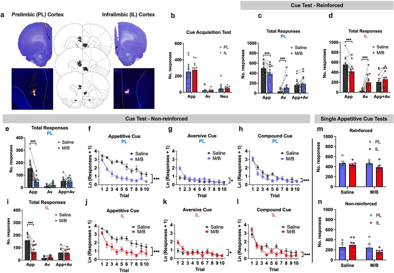
Systems/Circuits
Cortico-Striatal Control over Adaptive Goal-Directed Responding Elicited by Cues Signaling Sucrose Reward or Punishment
Animals readily learn to identify sensory stimuli that signal the opportunity for reward or the threat of injury and to make appropriate responses when such cues are present. Animals can also learn to suppress actions when conflicting cues are present or when a particular cue is no longer a reliable predictor of an outcome. The ability of sensory information to drive appropriate behavior depends partly on the prelimbic (PL) and infralimbic (IL) areas of the medial prefrontal cortex and their respective targets in the core and shell regions of the nucleus accumbens (NAc). Much work has suggested that PL promotes approach or avoidance responses to cues signaling reward or punishment, whereas IL inhibits these responses when the cues lose predictive value. Hamel et al. extended this work with evidence that both L and IL regulate behavior when conflicting cues are present. Rats learned to press a lever to receive sucrose when a light flashed and to withhold lever presses to avoid shock when a tone played. After training, inactivating either PL or IL reduced lever pressing in response to the light cue and increased lever pressing in response to the sound cue, regardless of whether the sucrose or shock continued to be delivered. This suggests rats were less able to choose the appropriate action in response to conflicting cues.

Behavioral/Cognitive
Contingent Amygdala Inputs Trigger Heterosynaptic LTP at Hippocampus-To-Accumbens Synapses
After an animal learns to associate a particular sensory stimulus with a reward or threat, the animal will be motivated to make appropriate behavioral responses when it encounters the stimulus in the future. The ability of cues to motivate behavior depends on medium spiny neurons in the nucleus accumbens. These neurons receive convergent input from the basolateral amygdala, which conveys information about valence states, and the ventral hippocampus, which conveys contextual information. By using optogenetic techniques to activate these inputs in brain slices, Yu et al. found that co-activation caused long-term potentiation of hippocampal inputs without affecting amygdala inputs. The effects required the activation of D1-type dopamine receptors. Thus, this work suggests a plausible mechanism by which neutral cues may acquire the ability to motivate action.

Behavioral/Cognitive
Distinct Progressions of Neuronal Activity Changes Underlie the Formation and Consolidation of a Gustatory Associative Memory
In gustatory cortex, tastes are represented by distinct spatiotemporal patterns of activity across ensembles of broadly tuned neurons. This population activity encodes both the identity and the valence of tastes. Therefore, ensemble activity is altered during the development of conditioned taste aversion (CTA). Because this learning is mediated by changes in synaptic strength between individual cells in ensembles, Arieli et al. how changes at the single-cell level relate to changes in population dynamics. Single-unit recordings revealed that individual neurons' responses changed in different directions (increases and decreases) and at different times to produce an overall increase in firing during the acquisition and consolidation phases of CTA. The work suggests that taste learning involves continuous adjustments in the activity of individual neurons to achieve the desired changes in network state.
Listen to Anan Moran and Elor Arieli discuss their paper on Episode 20 of Neuro Current: An SfN Journals Podcast.
Behavioral/Cognitive
Oxytocin and the Punitive Hub—Dynamic Spread of Cooperation in Human Social Networks
Cooperation among unrelated individuals is essential for large societies to function. To ensure cooperation, groups often punish freeloaders. Remarkably, oxytocin, a neuropeptide produced by the hypothalamus not only increases people’s willingness to cooperate, but also makes them more likely to punish those who fail to cooperate. Using economic games and computer simulations, Li et al. found that administering oxytocin to a few well-connected people in a group increased cooperation throughout the group. Surprisingly this effect did not stem from an increase in willingness to cooperate, per se, but rather from increasing the tendency to punish those who acted selfishly.

Neurobiology of Disease
STAT1 Contributes to Microglial/Macrophage Inflammation and Neurological Dysfunction in a Mouse Model of Traumatic Brain Injury
After traumatic brain injury (TBI), microglia and macrophages become activated in various ways to promote inflammation and tissue repair. Although initially beneficial, proinflammatory microglia and macrophages can cause secondary tissue damage and worsen recovery prospects. Therefore, selectively reducing the number of proinflammatory cells may improve outcomes after TBI. Zhao, Ma, et al. did this by administering fludarabine, which inhibits STAT1, a transcription factor that promotes proinflammatory phenotypes in peripheral macrophages and is upregulated in the brain after traumatic injury. Administering fludarabine after TBI in mice reduced the number of proinflammatory microglia and macrophages in the brain and increased the number of prorepair cells. It also reduced brain levels of inflammatory cytokines, tissue loss, and axonal damage, and it sped recovery of sensorimotor function. These findings suggest that fludarabine may improve functional recovery in patients with traumatic brain injury.